
Messy life-producing cellular process caught in action for first time




All living things have a fleet of tiny copy machines that turn instructions from DNA into RNA, the molecules responsible for helping build proteins that keep organisms alive. One of those nanoscale machines, called eukaryotic RNA polymerase II, performs a crucial early step required by nearly every biological process. It makes messages, copying and carrying instructions from the cell nucleus to make proteins.
Until recently, scientists had only witnessed the eukaryotic RNA at work in carefully assembled test tubes, stripped of the chaotic realities of life inside a cell. Now, a team led by Penn State researchers has captured a glimpse of this molecular machinery as it operates inside living organisms. They reported their findings in the journal Nature Communications.
“This is the first time we’re seeing this process as it actually happens; the way it acts when no one is watching,” said Katsuhiko Murakami, the Stanley Person Professor of Molecular Biology and director of the Huck Center for Structural Biology at Penn State and co-corresponding author on the study. “Previously, we had to use highly purified samples under ideal lab conditions to visualize their structures, which is not how life really works. Now, we must completely change our thinking because what we’re seeing is not anything we have seen before.”
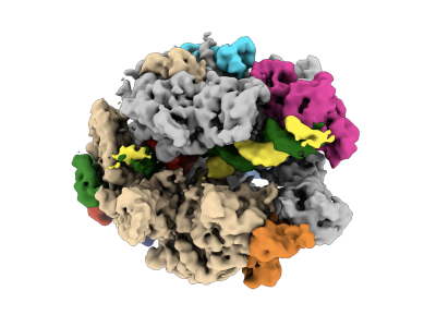
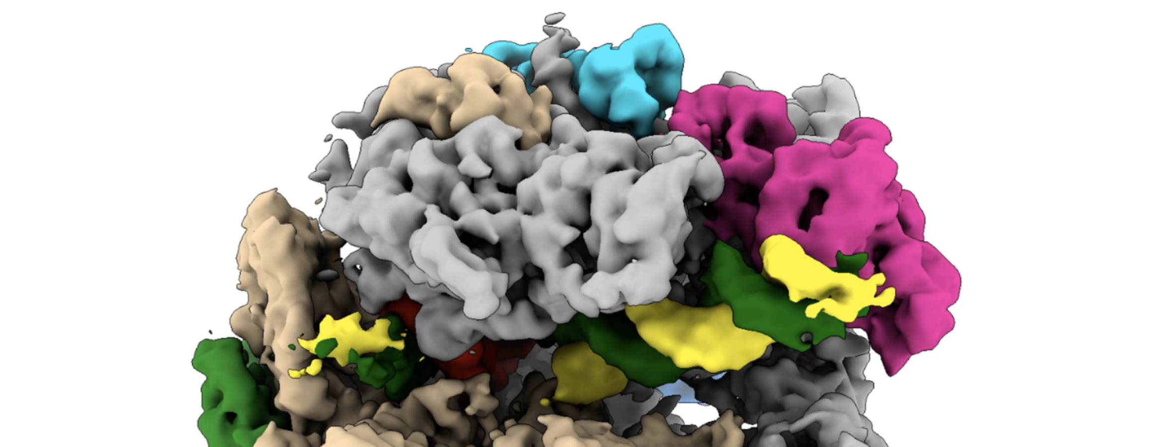
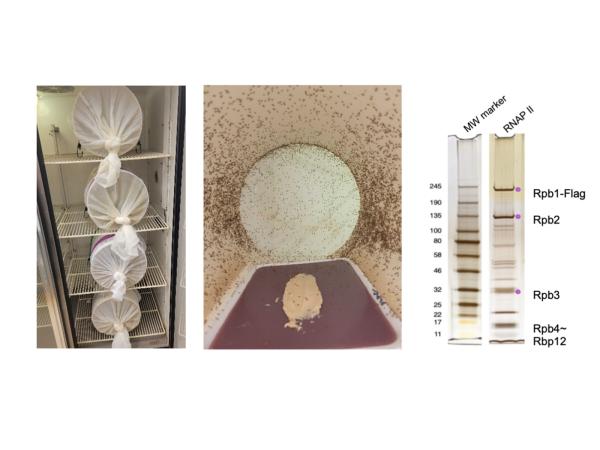
Using fruit fly embryos, the team developed a method to extract intact “transcription complexes,” the clusters of RNA polymerase II and DNA involved in reading and copying genes. They then used cryo-electron microscopy (cryo-EM), a powerful imaging technique that freezes molecules in place and visualizes them at near-atomic detail, to map what they found. What emerged was a far more dynamic and surprising picture of the copy machine process, called gene transcription, than they had expected.
Murakami explained that the project began in 2021 when David Gilmour, emeritus professor of biochemistry and molecular biology at Penn State, showed him purified RNA polymerase II extracted from a fruit fly embryo.
“It wasn’t clean, but it sparked an idea,” he said. “We could use cryo-EM to analyze native transcription complexes from it. After years of hard work, we’ve now captured these complexes in a near-native state and what we found was actually pretty surprising.”
Prior to this study, RNA polymerase II was thought to be made up of 12 subunits that come together to form one complete unit, so most researchers assumed they all looked the same in cells, he said. But the team’s results showed that’s not always the case. Some of them have all 12 subunits, like expected, but others are missing two subunits leaving them with only 10 subunits.
“That kind of variation wasn’t what people thought would happen, so it was a really unexpected finding,” Murakami said.
The discovery may offer clues to how cells manage the delicate balance between tightly packing DNA and making it accessible when needed. Such moments of transition are difficult to observe in traditional experiments, he said, making their appearance in the team’s cryo-EM imaging study particularly significant.
“From a basic science perspective, the approach used to be pretty straightforward,” said Jean-Paul Armache, assistant professor of biochemistry and molecular biology at Penn State and co-author of the paper. “Researchers would try to capture one clear picture of something, study what they saw and then publish their findings. Now, instead of looking at just one, controlled version, we can study a mix at the same time and understand how they vary. That gives us a much fuller, messier and more accurate picture of what’s going on. We’re not seeing one, sanitized process; it’s everything simultaneously — how life really happens.”
The study is part of a broader shift in modern biology, Armache said, of researchers moving away from laboratory-built systems toward observing molecules as they exist in living cells. A better understanding of how the gene-reading process works in real-life conditions offers a more accurate blueprint of how cells function, which can have applications for fields like medicine, he said.
The team’s approach could open the door to studying many other complex cellular processes in their natural environments, Murakami said.
“We’re starting to use this system more broadly, not just in one kind of organism, but also in others like archaea, which are part of the microbiota of all organisms,” Murakami said. “We’re trying to understand how these molecules behave in real conditions and different environments, so this is just the beginning for what we’ll be able to see.”
Other authors on the study include Paul Babitzke, the Don Bryant Chair in Microbial Physiology at Penn State; Ganesh Anand, associate professor of chemistry, and biochemistry and molecular biology at Penn State; Natalie L. Venette-Smith, previously a doctoral degree candidate in Molecular, Cellular, and Integrative Biosciences program at Penn State now at UT Southwestern Medical Center; Varun Venkatakrishnan, a doctoral degree candidate in chemistry at Penn State; Rishi K. Vishwakarma previously an assistant research professor of biochemistry and molecular biology at Penn State and now at St. Jude Children’s Research Hospital; Roberta Dollinger, previously a doctoral degree candidate in Biochemistry, Microbiology and Molecular Biology program of Penn State and now at the University of Wisconsin–Madison; Josie Schultz, previously a bachelor degree candidate in Penn State’s Department of Biochemistry and Molecular Biology and now at the Frederick National Laboratory for Cancer Research.
This work was supported by the Pennsylvania Department of Health and the National Institutes of Health under award numbers R35 GM156623, R21AI168948, R01 GM047477, R01 GM098399 and R35 GM161751. The content is solely the responsibility of the authors and does not necessarily represent the official views of the National Institutes of Health.